An Automatically Operating Microscope
Basic part of the Microscopic Image Analysis System (MIAS ® )
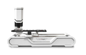
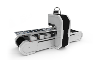
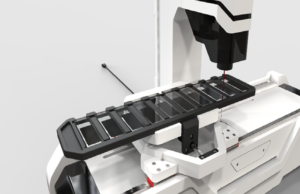
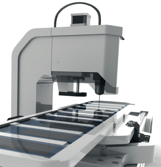
It is an automated light microscope,
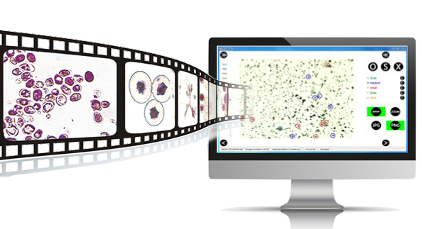
designed to make series of images
from separate samples.
DIGITIZE YOUR SAMPLES
This way samples are digitized as an ordered series of images at high
resolution.
This digitalization provides the basis of:
- Sample evaluation separate from microscope and lab
- An archive for documentation and report
- An archive for documentation and report
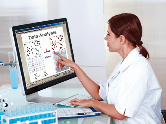
- Automatic image analysis for selected topics.
FEATURES OF THE OPERATION
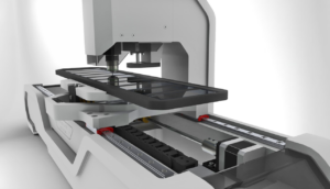
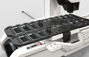
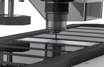
This apparatus consists of a microscopic system with high resolution
optic and a large cross table. The mechanics are designed to operate
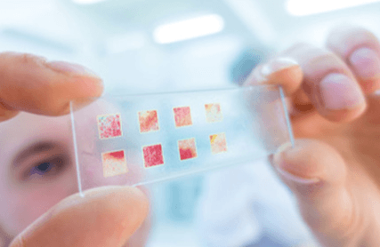
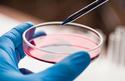
similar to the routine eye inspection of appropriate standard samples
in light microscopy.
The aeroScope ® is connected to a PC with specific software. After easy
setting of the parameters for each sample, the aeroScope ® can be
started to scan automatically up to ten different slides and make
appropriate series of images.
The location of each image on the slide and the parameters set for
the scanning of the appropriate sample are carefully documented.
The image files and corresponding data will be saved in a file system
generating a digital archive of the samples.
MAIN BENEFITS
The digitalization of samples offers a number of opportunities:
- The automation facilitates a standardization of the process of the sample analysis.
- The evaluation of the images is independent from the microscope and a laboratory. It can be done at a different location, e.g. from home when the archive is made accessible there.
- The recognized particles of interest can be easily related to the time of sampling by the positions of the images.
- The digitalization of the samples allows a repeated, extended and sophisticated analysis.
Custom solutions
The development and assembling of all components of the aeroScope ® is in responsibility of our company. Therefore
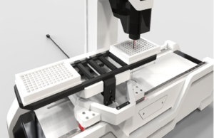
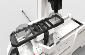
- The mechanical setup can be altered to related applications with other sample holders, illumination, magnification and camera, and
- All software can be adapted to customer’s requirements, because it is made in house.
